Ciliated preadipocytes are sited on blood vessels in fat pads. DNA is blue, vasculature is white, lipid droplets in mature fat cells is green, centrosome is green, and ciliary axoneme is red. Murine white adipose tissue. Adapted from Hilgendorf et al. Cell (2019).

Mouse cell line model and primary isolated mouse and human preadipocytes are ciliated. DNA is blue, centrosome is green, and ciliary axoneme is red. Scale bar is 5µm. Adapted from Hilgendorf et al. Cell (2019).

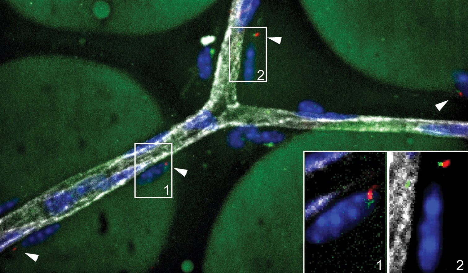
Ciliated preadipocytes are dotted along blood vessels in fat pads. DNA is blue, vasculature is white, centrosome is green, and ciliary axoneme is red. Murine mesenteric white adipose tissue. Adapted from Hilgendorf et al. Cell (2019).

Fat pads expand by generating more fat cells and storing additional fat in existing fat cells. Balancing production of fat cells with lipid accumulation is critical for “healthy” fat. This micrograph shows a mouse gonadal fat pad with mature fat cells visualized by phase microscopy and a population of ciliated fat progenitor cells (cilia in red and centrioles in green) located along the vasculature.